June 2026 – Dr. Brahim Mehadji Receives STAIR HEALTH Proof-of-Concept Grant for PET-Guided Cancer Surgery

Dr. Brahim Mehadji received a UC Davis STAIR HEALTH Proof-of-Concept Grant in support of his project, Precision PET Probe: Revolutionizing Minimally Invasive Cancer Surgery.
Dr. Mehadji's work aims to develop a compact PET probe designed to guide surgeons during minimally invasive cancer removal procedures. Currently, one of the key challenges in cancer surgery is confirming in real time that all tumor tissue has been removed. Dr. Mehadji's probe would bring PET radiotracer imaging directly into the operating room, giving surgeons a precise, live view of tumor boundaries during the procedure itself. A major hurdle this project addresses is the current lack of laparoscopic devices capable of detecting PET tracers, making this a genuinely novel contribution to the field.
The one-year grant will fund prototype development and early validation studies, helping move this technology closer to clinical use. Dr. Mehadji was one of only three recipients selected across UC Davis this cycle.
The STAIR HEALTH program invests in translational research with clear potential to improve patient care and accelerate the path from laboratory discovery to real-world clinical impact.
Congratulations, Brahim, on this exciting recognition!
June 2026 – Dr. Lorenzo Nardo Receives the Drs. Jane and Abass Alavi Mars Shot Research Award at SNMMI 2026

The EXPLORER Molecular Imaging Center is delighted to congratulate Dr. Lorenzo Nardo, Professor of Radiology at UC Davis Health, on receiving the Drs. Jane and Abass Alavi Mars Shot Research Award at the 2026 SNMMI Annual Meeting. The proposal, Zirconium-89 Certolizumab PET Imaging in Patients with Rheumatoid Arthritis, was selected to receive a grant of up to $110,000 from the SNMMI Mars Shot Research Fund, which is focused on advancing molecular imaging research in multiple domains including infection and inflammation.
Rheumatoid arthritis (RA) is a chronic autoimmune condition where the immune system attacks the joints, causing painful inflammation and, over time, irreversible joint damage. Many patients are treated with TNF- blocking medications, which work by neutralizing a key inflammatory protein that drives joint destruction. Certolizumab pegol (Cimzia®) is one such drug, and its unique molecular struture makes it well-suited to be tagged with a radioactive label for PET imaging without changing how it behaves in the body.
The ⁸⁹Zr-labeled certolizumab tracer was originally developed by Dr. Henry VanBrocklin, Professor of Radiology and Biomedical Imaging and Director of Radiopharmaceutical Research at UCSF, whose lab has pioneered the labeling of antibody-based drugs with zirconium-89 for immunoPET imaging across several disease areas including rheumatoid arthritis. This study brings the tracer to UC Davis in a collaborative effort co-led by Dr. Barton Wise, Professor of Rheumatology and Orthopedic Surgery, whose clinical expertise in arthritis will be integral to patient selection and outcome assessment.
By using the drug itself as a PET tracer, doctors can see exactly where it travels and accumulates in inflamed joints, potentially predicting whether a patient is likely to respond to treatment before committing to a prolonged course of therapy. This kind of precision could spare patients from ineffective treatments and guide more informed clinical decisions.
Congratulations, Lorenzo and team, on this well-deserved recognition!
June 2026 – Dr. Yansong Zhu Receives First Place Young Investigator Award at SNMMI 2026

Dr. Yansong Zhu has been honored with the First Place PIDSC Young Investigator Award at the 2026 SNMMI Annual Meeting, the premier annual gathering of nuclear medicine and molecular imaging professionals worldwide. Yansong's work, "Super-resolution PET-enabled dual-energy CT imaging with application to different radiotracers," was selected as one of six finalists from a competitive pool of nearly 300 submitted abstracts and ultimately received the top distinction.
The study, conducted using UC Davis's uEXPLORER Total-Body PET/CT scanner, introduces a novel method for getting more informative images from existing PET/CT scanners, without requiring any additional hardware or extra radiation exposure to the patient. Normally, PET scans produce images that are much blurrier than CT scans, making it harder to distinguish fine tissue details. Yansong's approach derives a second gamma-ray CT image entirely from the PET scan's own data, bringing it up towards CT-level clarity, aided by advanced AI-driven reconstruction method.
Beyond sharper images, this technique also enables a richer view of tissue composition: by combining information from two different CT energy levels (the standard x-ray CT and the higher-energy PET-derived CT), clinicians can better distinguish between bone, soft tissue, and other materials in a single scan session. The team demonstrated that the method works reliably across different types of radiotracers, expanding its potential use across a wide range of clinical scenarios, from cancer imaging to body composition and bone health assessment.
Congratulations to Yansong and the entire team!
Related Link:
May 2026 – Professor Simon Cherry Elected Fellow of the Royal Society

The EXPLORER Molecular Imaging Center is proud to celebrate a landmark achievement: Professor Simon R. Cherry has been elected a Fellow of the Royal Society (FRS), one of the most prestigious scientific honors in the world. The announcement was made on May 27, with only 90 scientists across the globe receiving the honor this year.
Founded more than 360 years ago, the Royal Society is a self-governing Fellowship made up of many of the world's most eminent scientists, engineers, and technologists — a community whose past members include Isaac Newton and Charles Darwin. Election to Fellow is recognition reserved for those who have made exceptional contributions to the advancement of science.
The Royal Society elected Cherry in recognition of his seminal contributions to biomedical imaging. Cherry has transformed imaging science across a more than 35-year career, including co-leading the development of EXPLORER, the world's first total-body PET scanner, alongside Professor Ramsey Badawi, a technology that has reshaped biomedical research and clinical medicine by enabling unparalleled insights into healthy and disease states in the human body. Cherry’s broader body of work spans detector and system development, PET/MRI hybrid imaging, Cerenkov radiation imaging, and quantitative analysis methods, with applications ranging from cancer and neurology to infectious disease and metabolism.
"I am incredibly honored to receive this recognition from one of the world's most prestigious and famous scientific institutions," Cherry said. "I feel very fortunate to have such a wonderful career, doing things that I love and surrounded by so many bright and generous people."
Congratulations, Simon, on this extraordinary and richly deserved recognition!
Related Links:
- Royal Society Fellow profile — https://royalsociety.org/people/simon-cherry-38141/
- UC Davis News — https://www.ucdavis.edu/news/uc-davis-medical-imaging-expert-elected-fellow-royal-society
May 2026 - Simon Cherry leads opening ceremony at SNMMI 2026

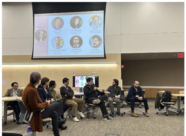
Our own Prof. Simon Cherry will lead the opening ceremony at the SNMMI meeting 2026. Simon will present exciting news in the field in his 15-min keynote lecture right before moderating an engaging panel discussion on Nuclear Medicine Access and Equity, joined by six representatives from both research and industry. One of the panelists will be our colleague Brad Schmidt, CEO of the Northern California PET imaging center, who we share our building with.
The schedule also contains an exciting discussion with the British Nuclear Medicine Society (BNMS) President Sabina Dizdarevic MD, PhD, who will afterwards present latest research and development from the BNMS in a highlight country video. The program highlights of this year’s SNMMI meeting will then be presented by Steve Cho, MD, Chair of Nuclear Medicine at MD Anderson. Finally, after some closing remarks by Simon, the attendees will proceed to the opening ceremony with the industry exhibitors for some light dinner and refreshments.
This year the society of nuclear medicine and medical imaging (SNMMI) holds their annual gathering in Los Angeles. According to the event website, "the 2026 SNMMI Annual Meeting will feature groundbreaking research and clinical innovation”. The goal is again to unite physicians, technologists, pharmacists, laboratory professionals, scientists, and industry to demonstrate latest developments that help pave the way towards better patient care.
Congratulations, Simon, for being invited to open such a distinguished event in our field!
May 2026 - Dr. Siqi Li Receives His First NIH R01 Grant
We are pleased to share that Dr. Siqi Li, Assistant Project Scientist in the lab of Dr. Guobao Wang, has received his first NIH R01 award to develop practicalPET parametric imaging methods for pediatric cancer imaging. The project focuses on advancing dynamic PET imaging techniques that provide more quantitative measures beyond conventional standardized uptake values (SUVs), without requiring lengthy scan times that are impractical for children.
The research will develop and evaluate a streamlined kinetic modeling approach, Relative Patlak plot, which can be directly applied to total-body clinical scans for parametric imaging without changing the existing scan protocol. Unlike other simplified methods that rely on the population-based database, this approach can adapt to different scanning time, protocols, and other tracers. The project will investigate applications for improved lesion detection, treatment response prediction, and broader implementation on both total-body and conventional PET scanners. By enabling efficient and quantitative whole-body PET imaging in children, the work aims to fill an important gap in pediatric cancer imaging and expand access to advanced PET methodologies previously limited largely to adult studies.
This award is an important milestone for Dr. Li and advances our lab’s broader efforts in quantitative molecular imaging. Congratulations to Siqi on this exciting achievement!
May 2026 - Xiaoyu Duan and Kevin Chung receive JNM’s Alavi-Mendell Awards for Best Publication

PhD candidate Xiaoyu Duan and postdoc Dr. Kevin Chung (now Instructor at MGH) received the Alavi-Mandell Award from the Journal of Nuclear Medicine (JNM) for their JNM papers published in 2025. The award was established by the families of Dr. Abass Alavi, MD and Dr. Gerald Mandell, MD to encourage young physicians and scientists to pursue a career in academic and research in nuclear medicine. Congratulations to both of our young scientists for their excellent contributions to our research field!
The corresponding publications are listed below:
Duan X, Sarkar S, Lyo V, Romeo S, Spencer BA, Matsukuma KE, Medici V, Corwin MT, Badawi RD, and Wang GB.
Quantification of 18F-FDG delivery rate for liver inflammation using shortened dynamic PET imaging protocols.
Journal of Nuclear Medicine, 66(11): 1834-1841, 2025.
Xiaoyu is jointly mentored by Dr. Guobao Wang and Dr. Ramsey Badawi.
Chung KJ, Chaudhari AJ, Nardo L, Jones T, Chen MS, Badawi RD, Cherry SR, Wang GB.
Quantitative total-body imaging of blood flow with high temporal resolution early dynamic 18F-Fluorodeoxyglucose PET kinetic modeling.
Journal of Nuclear Medicine, 66(6): 973-980, 2025.
Kevin was jointly mentored by Dr. Guobao Wang and Dr. Simon Cherry.
April 2026 - Best paper awards for Manish Das and Reimund Bayerlein

On April 15, the Editorial Board and Scientific Council of Bio-Algorithms and Med-Systems have announced that the Best Scientific Paper Award for 2024 has been presented to the article: “Development of correction techniques for the J-PET scanner” prepared by an international research team led by Manish Das and Reimund Bayerlein, et al. The Best Paper Award initiative was established in 2022 with the goal to recognize the importance of impactful and high-quality scientific contributions. Each year, the distinction is granted following a detailed evaluation of the scientific merit of the published works, their contribution to research advancement, and their relevance and visibility within the scientific community.
The awarded publication presents advanced correction methodologies for PET imaging data acquired using the modular J-PET scanner developed at the Jagiellonian university of Krakow. The study successfully demonstrates attenuation correction, randoms correction, and scatter correction and exhibits significant improvements in image quality. The results confirm the strong scientific and technological potential of plastic-scintillator-based J-PET systems in the development of more accessible PET diagnostics and future total-body PET imaging solutions. The editorial board called the publication a “remarkable achievement” and thanked the authors “for their valuable contribution to the advancement of medical imaging technologies”.
Congratulations Manish and Reimund for this important scientific contribution and the distinguished award!
April 2026 - Simon Cherry Receives ESMI Award for His Long-Term Contributions to Imaging Science

Dr. Simon R Cherry Distinguished Research Professor of biomedical engineering and radiology at the University of California, Davis, has received the 2026 ESMI Award from the European Society for Molecular Imaging (ESMI). This honor is among the most prestigious handed out in imaging science, celebrating a researcher for outstanding long-term contributions to the field.
Simon has transformed imaging science across his more than 35-year career. One of his many achievements is his massive contribution to co-developing (with his long-time friend and collaborator Ramsey Badawi) the EXPLORER – the world’s first total body PET scanner. This high-sensitivity imaging device constitutes a paradigm shift in nuclear imaging, biomedical research and has improved clinical practices.
In the ESMI’s award citation Dr. Cherry is recognized for his “outstanding scientific contributions to the field and for his unwavering perseverance, passion and visionary thinking”. He is furthermore considered an “outstanding, open-minded, curious scientist”. Needless to say that we couldn’t agree more with the ESMI award committee! Upon receiving the ESMI award, Simon expressed his deep appreciation, attributing the success to the incredible contributions of his past and present lab members and collaborators. We are excited to see the next great idea shaping the landscape of medical imaging in nuclear medicine!
Congratulations, Simon!
April 2026 - Reimund Bayerlein-Awarded NIH R03 Grant for Exploring the Potential of Sparse Total-Body PET

Dr. Reimund Bayerlein, an Assistant Project Scientist based at University of California, Davis, has been awarded a 2-year NIH R03 research grant as Principal Investigator to explore cost-efficient sparse total-body positron emission tomography (PET) imaging and its impact on quantitative image quality - particularly in dynamic acquisition modes. This research project focuses on the development of novel system designs to improve accessibility and performance. Using data acquired on the uEXPLORER total-body PET/CT scanner, the work simulates cost-efficient scanner configurations by artificially downsampling detector data to mimic sparse acquisitions prior to image reconstruction. This approach enables a rigorous, data-driven evaluation of how reduced system complexity affects image quality and quantitative accuracy, without requiring physical hardware modifications. By identifying sparse system configurations that preserve key performance characteristics, this research aims to considerably reduce system cost while maintaining clinical and research utility. Ultimately, this could improve access to high-performance PET imaging, enabling broader use of quantitative and dynamic imaging techniques in both clinical practice and translational research.
The project will be conducted at the EXPLORER molecular imaging center (EMIC) at UC Davis, with total funding of $161,000 over 2 years from the National Institute of Biomedical Imaging and Bioengineering (NIBIB). This award marks an important milestone in Dr. Bayerlein’s research career and represents a significant step forward in advancing affordable, high-sensitivity total-body PET imaging.
Congratulations, Reimund!
March 2026 - Xiaoyu Duan: SNMMI Bradley-Alavi Fellowship

Xiaoyu Duan, a PhD candidate in the Wang lab, who has received the Bradley-Alavi Fellowship from the Society of Nuclear Medicine and Molecular Imaging. This fellowship supports outstanding trainees pursuing research in molecular imaging and nuclear medicine.
Xiaoyu’s fellowship project focuses on developing quantitative PET methods for assessing liver inflammation in metabolic dysfunction-associated steatotic liver disease (MASLD). Her work aims to improve dynamic PET imaging protocols and extend kinetic modeling to multi-organ analysis using total-body PET, enabling more practical and informative imaging biomarkers for MASLD patients.
Xiaoyu previously published first-author work in the Journal of Nuclear Medicine 2025 and received the Runner-Up Best Student Paper Award at the IEEE Medical Imaging Conference 2024. This fellowship will support her continued research in parametric PET.
Xiaoyu is jointly supervised by Dr. Guobao Wang and Dr. Ramsey D. Badawi.
Congratulations Xiaoyu!
February 2026 - Liu Guobing: Star of Medical Ethics and Professional
Excellence

Liu Guobing, MD, is an Attending Physician in the Department of Nuclear Medicine at Zhongshan Hospital, Fudan University. He serves as a Youth Committee Member of the 6th Committee of the Nuclear Medicine Branch of the Chinese Society of Medical Imaging Technology and as a member of the Imaging Group of the Nuclear Medicine and Molecular Imaging Committee under the 11th Council of the Shanghai Medical Association.
He is engaged in nuclear medicine diagnosis and treatment as well as molecular imaging research. As first or corresponding author, he has published more than 50 academic papers, including over 40 SCI-indexed papers, with 7 papers published in top-tier (CAS Zone 1) journals. He has served as editor-in-chief of one Chinese and one English monograph, associate editor of one Chinese monograph, and contributor to one English monograph.
He has led multiple research projects including National Natural Science Foundation of China grants, subprojects of key national R&D programs from the Ministry of Science and Technology, and several provincial, municipal, and talent-development programs. He has delivered more than 10 oral presentations at major domestic and international academic conferences. He has participated in six patent applications and has received the New Technology Application Promotion Award of Zhongshan Hospital (Fudan University) four times as a key contributor.
November 2025 - School or Medicine Research Rock Star Awards for Kevin Chung and Phu Huynh


We are thrilled to share that the UC Davis School of Medicine Office of Research honored two outstanding members of our EMIC team at the recent University of California, Davis - School of Medicine Research Celebration. Two of the five Research Rock Star Awards were awarded to Phu Huynh – Nuclear Medicine Technologist Supervisor II and Kevin Chung, Postdoctoral Fellow with the Wang Lab.

Phu Huynh oversees all studies using the EXPLORER Total-Body PET/CT scanner – supporting more than 30 research programs spanning cancer, autoimmune, neurologic, and metabolic diseases. He is at the operational center of clinical research at EMIC, developing and implementing advanced imaging protocols, to ensuring regulatory compliance, equipment procurement/maintenance. He embodies reliability, adaptability, technical acumen and a deep commitment to the science and the people behind it.
Dr. Kevin Chung’s groundbreaking work leverages the EXPLORER total-body PET/CT system to study how the blood–brain barrier (BBB) changes in early disease. By developing new imaging techniques to quantify BBB permeability, his research provides crucial insights into how systemic and neurologic processes interact long before symptoms appear. His findings are helping to redefine how we detect, monitor, and understand neurodegenerative and inflammatory disorders at their earliest stages. This research has gained international recognition, earning Dr. Chung several prestigious awards and positioning him as a rising leader in molecular imaging.
Congratulations again, Phu and Kevin!
November 2025 - Dr. Guobao Wang wins IEEE Medical Imaging Technical Achievement Award

Congratulations to Guobao Wang on receiving the IEEE Freek J. Beekman Medical Imaging Technical Achievement Award at this year's IEEE NSS/MIC/RTSD conference! This award is given annually (previously only in in even-numbered years)to a mid-career individual in recognition of significant and innovative technical contributions to the field of medical imaging science. The award, consisting of $2,000, a certificate, and a plaque, is open to all active members of the medical imaging community and is presented at the IEEE Medical Imaging Conference. This year, this annual meeting took place in Yokohama, Japan, in November.
Dr. Wang was honored for his contributions to advanced image reconstruction and translational kinetic modeling. We are happy that his innovative work for PET imaging has been recognized!
October 2025 - New AI Model “AMDiff” Boosts PET Image Quality and Lesion Segmentation

UC Davis collaborators at Yale University have developed AMDiff, a groundbreaking artificial intelligence model that simultaneously cleans up noisy PET scans and identifies organs and lesions – tasks traditionally handled separately. By combining these functions into a unified framework, AMDiff takes advantage of the natural link between image clarity and anatomical accuracy.
Yale postdoc Menghua Xia developed AMDiff using advanced diffusion-based denoising mechanism and novel nnMamba segmentation architecture. AMDiff produces clearer images and more precise measurements, even from scans with very low tracer counts. In multi-center tests, the AMDiff showed over 20% lower image error and notably higher lesion segmentation accuracy than the baseline single-task methods.
The model also enables computation of key clinical metrics like total lesion glycolysis directly from low-dose scans, potentially reducing radiation exposure and improving diagnostic confidence across diverse imaging systems.
October 2025 - Excellent performance of EMIC researchers at Radiology Research Symposium


Two of our young research members were awarded with the first prize for the best abstract contribution to this year’s Radiology Research Symposium held at the Betty Irene More Hall on October 22. David Melnichuk won in the category of best undergraduate submission for his investigations of lesions detectability of PET images that were denosied with artificial intelligence tools. Furthermore, Shivani took the prize for the best post- doctoral contribution for her evaluation of 18F-FAPI-74 total-body PET as a quantitative biomarker for fibrosis staging in Metabolic Dysfunction-Associated Steatotic Liver Disease (MASLD). Congratulations to both winners!
Once a year, the Department of Radiology organizes a symposium to show the current research projects through oral present tions and a poster exhibition. This year, a novel Shark Tank session was included in the program where young researchers could pitch their ideas to a panel of experts. Two of our EMIC researchers (Brahim Mehadji and Reimund Bayerlein) were amongst the finalists. The $50,000 award was eventually handed to Michael Larson to fund his research on a novel suture delivery system e abling ultrasound- guided hernia and prolapse repair. Congratulations!
October 2025 - Total-Body PET/CT Could Cut Radiation Dose for Lymphoma Patients
A new study by Clemens Mingels from Bern, Switzerland, suggests that total-body (TB) PET/CT scans may allow for much lower radiation doses without compromising diagnostic accuracy in lymphoma patients. Researchers tested how far injected activity of the tracer
¹⁸F-FDG could be reduced while still providing reliable images for therapy response assessment.
Twenty-four patients were scanned at 1 and 2 hours post-injection using both standard and simulated low-dose settings. Even when tracer doses were cut to as little as 1/12th of the standard level, key tumor measurements – including metabolic tumor volume and lesion glycolysis – remained accurate. Importantly, therapy response classifications (Deauville scores) were unchanged down to 0.25 MBq/kg at 1 hour and 1.0 MBq/kg at 2 hours.
These results indicate that TB PET/CT can maintain clinical reliability with greatly reduced tracer doses, potentially improving patient comfort and safety during cancer monitoring.
October 2025 - New AI Tool “PUMA” Enhances PET/CT Imaging with Multitracer Fusion
In a collaboration with UC Davis Lalith Sundar – a researcher based in Munich, Germany – has unveiled a powerful new imaging tool called PUMA (PET Unified Multitracer Alignment) that allows doctors and scientists to combine multiple PET/CT scans into a single, unified view. Traditionally, PET/CT imaging uses one tracer at a time, limiting how much information can be gathered about complex diseases. PUMA overcomes this by aligning scans from different tracers using advanced artificial intelligence and precise image registration.
In a study involving 114 patients from multiple centers, PUMA achieved highly accurate organ alignment and preserved key imaging data, ensuring no loss of diagnostic quality. The tool can even visualize up to three tracers simultaneously using red–green–blue color channels.
As an open-source framework, PUMA provides a vendor-independent way to enrich molecular imaging without changing existing clinical protocols – paving the way for more comprehensive disease characterization and improved treatment planning.
October 2025 - Dr. Negar Omidvari Joins the Department of Radiology as an Assistant Professor
Great news! The UC Davis Department of Radiology has a new faculty member and it's none other than our very own Dr. Negar Omidvari. As of October 2025, she has joined the Department of Radiology as an Assistant Professor. Her research group — the Omidvari Molecular Imaging and Quantification (OMIQ) Laboratory — is focused on quantitative total-body PET imaging of the immune system, with three newly funded studies on long-COVID, non-small cell lung cancer, and Alzheimer’s disease that use dynamic imaging of the [18F]F-AraG tracer for evaluating T cell activation and beyond! We congratulate her on the new position! We look forward to her exciting contributions to our research field.
October 2025 - Dr. Negar Omidvari Selected as a Semifinalist Candidate in Minnies 2025 Award for Most Influential Radiology Researcher
Congratulations to Dr. Negar Omidvari for being selected as a semifinalist for Most Influential Radiology Researcher award by AuntMinnie.com's campaign to recognize the best and brightest in medical imaging. This year, 18 candidates were selected as semifinalists in the Most Influential Radiology Researcher for the period of September 1, 2024, to September 1, 2025.
The Minnies awards have recognized excellence in radiology since 2000. The semifinalist list is compiled annually based on nominations from members of AuntMinnie.com. Winners are selected by an expert panel in two rounds of voting. The finalists are announced in late October, followed by the winners in early November.
August 2025 – Dr. Guobao Wang and his collaborators at UC Davis have secured a $2.5 million NIH grant to advance a novel hybrid imaging technique combining PET and dual- energy CT, promising better detection of cancer, bone, and heart diseases.
The four-year funding supports PET-enabled Dual-Energy CT, which uses PET data to generate high-energy CT images alongside the standard ones, revealing tissue composition without extra radiation or new CT hardware which is normally required for dual-energy CT imaging.
A paper in the European Journal of Nuclear Medicine and Molecular Imaging by Yansong Zhu et al. last year showcased this breakthrough using the innovative EXPLORER total- body PET scanner, proving its real-world potential
This new method could enable sharper distinction between healthy and cancerous tissues and enhance treatment response monitoring in a single scan. The method could also improve bone marrow images and offers new insights into heart disease such as inflammation. With refinement and fine tuning, this method could make advanced imaging more accessible worldwide without costly upgrades.
August 2025 - Dr. Negar Omidvari Awarded $3.2M NIH R01 Grant for Studying Long COVID
Congratulations to Dr. Negar Omidvari, Assistant Project Scientist in the Department of Biomedical Engineering, who has been awarded an R01 grant from the National Institute of Allergy and Infectious Diseases (NIAID) to investigate the immune and systemic manifestations of post-acute sequelae of SARS-CoV-2 infection (PASC, or “Long COVID”).
This four-year, $3.2 million collaborative project with UCSF will leverage the UCSF Long-term Impact of Infection with Novel Coronavirus (LIINC) clinical infrastructure for patient recruitment, symptom evaluation, and clinical phenotyping. Participants will then visit UC Davis’s EXPLORER Molecular Imaging Center (EMIC) for blood tests and total-body PET/CT imaging on the uEXPLORER scanner using the [18F]F-AraG radiotracer, which targets activated T cells. The study titled: "Multi Parametric Total-Body Imaging of Immune Activation in Post Acute Sequelae of SARS-CoV-2 (PASC)" will use advanced kinetic modeling to identify sites of immunological perturbation and vascular dysfunction in PASC patients, offering a total-body view of tissue-level manifestations of PASC.
August 2025 – A new study led by Dr. Clemens Mingels demonstrated that total-body PET/CT scans can safely reduce radiation doses for assessing lymphoma therapy response, potentially cutting exposure by up to 92% without compromising accuracy.
In a study published in the Journal of Nuclear Medicine, 24 lymphoma patients underwent [18F]FDG scans at standard 3 MBq/kg doses, with simulations testing reductions down to 0.125 MBq/kg. Doses as low as 0.25 MBq/kg at 1-hour post-injection and 1.0 MBq/kg at 2 hours maintained reliable tumor measurements and Deauville scores for treatment evaluation. Tumor metrics like SUVmax and metabolic volume showed no significant changes, though noise increased at ultra-low doses, risking false positives in response
assessment.
This advance, leveraging the scanner's high sensitivity, is especially beneficial for young patients, children, and pregnant women facing multiple scans and secondary cancer risks. Further trials are planned to validate real-world low-dose applications.
June 2025 - Simon Cherry gave the Steve Mark Larson Lecture in Advanced Nuclear Medicine
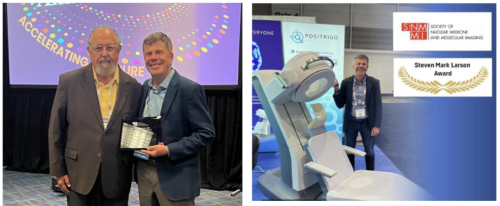
Congratulations to Prof. Simon Cherry for the honor of delivering the inaugural Steven Mark Larson, MD Lecture in Advanced Nuclear Medicine at 2025 Annual Meeting of the Society of Nuclear Medicine and Molecular Imaging (SNMMI). This new honor is among the most distinguished recognitions at SNMMI, celebrating outstanding contributions to the field of advanced nuclear medicine. Named for Dr. Steven Mark Larson, a pioneer in molecular imaging, the lecture highlights transformative research shaping the future of our field.
Prof. Cherry’s selection reflects his groundbreaking work in molecular imaging and his lasting impact on advancing nuclear medicine research and technology. We are proud to celebrate this achievement with the broader scientific community!
June 2025 – Ekaterina Shanina Received the 2024 Early Career Researcher Award from Physics in Medicine & Biology
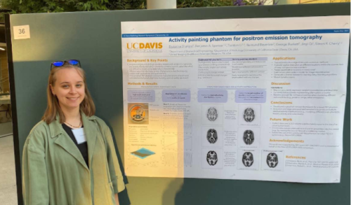
We’re proud to celebrate another superstar trainee: PhD student Ekaterina Shanina was recently awarded the 2024 Early Career Researcher Award from Physics in Medicine & Biology for her outstanding paper, “PICASSO: a universal brain phantom for positron emission tomography based on the activity painting technique”. Her paper was selected as the top contribution to the journal’s 2024 Focus Collection, recognizing innovative work in imaging science.
Her award-winning research introduced PICASSO—a leading-edge, dynamic brain phantom for PET imaging that sets a new standard for evaluating scanner performance. Unlike traditional phantoms, PICASSO eliminates cold walls and air bubbles that distort measurements and introduces a novel “activity painting” method to simulate complex, realistic distributions of radiotracer uptake. This allows researchers to better test and calibrate high-resolution PET systems using brain-like activity patterns that mimic real physiology.
Developed under the mentorship of Professors Simon Cherry and Jinyi Qi, PICASSO is already proving invaluable in evaluating systems like the NeuroEXPLORER, and it has wide-reaching potential to improve the accuracy and reliability of neuroimaging studies.
Congratulations, Katya!
Related Links:
- 🔗 See her news coverage on Physics World: https://physicsworld.com/a/phd-student-ekaterina-shanina-wins-early-career-researcher-award-for-pet-phantom-study/
- 🔎 You can read Katya’s paper at: https://doi.org/10.1088/1361-6560/ad84b5
- 👩🔬 Also, check out her work under “Poster Presentations” from the 2024 UC Davis Radiology Research Symposium. Katya gave a great e-presentation of her work: https://health.ucdavis.edu/radiology/research/symposium.html
June 2025 – Negar Omidvari received the Tracy Lynn Faber Memorial Award from the SNMMI PIDSC
Congratulations to Dr. Negar Omidvari, Assistant Project Scientist in the Department of Biomedical Engineering, for receiving the Tracy Lynn Faber Memorial Award from the Society of Nuclear Medicine and Molecular Imaging (SNMMI) Physics, Instrumentation and Data Sciences Council (PIDSC)! The Award is given either to an individual who has significantly promoted the advancement of women in medical imaging sciences, or to a woman in early- or mid-career who has made one or more significant contributions to medical imaging sciences. Dr. Omidvari received this award for her co tributions to molecular imaging science, including instrumentation, methodology and application to the immune system.
Dr. Omidvari is advised by Dr. Simon Cherry, Distinguished Research Professor in the Departments of Biomedical Engineering and Radiology.
June 2025 - Siqi Li received an Innovative Development Award from UC Davis Academic Federation

Congratulations to Dr. Siqi Li, assistant Project Scientist in the department of radiology, for receiving an Innovative Development Award from UC Davis Academic Federation. This award provides a pilot grant for Siqi to develop “Generalizable Deep Learning for Improving Pediatric PET Imaging.” He proposes developing a deep kernel model using a high-quality dataset collected from the ultra-high sensitivity total-body PET scanner EXPLORER. Once trained, this model can be used for tomographic image reconstruction across various scanners for low-dose
or fast pediatric PET imaging. Dr. Li is advised by Dr. Guobao Wang, Professor and Associate Vice Chair of Research in the Department of Radiology.
June 2025 – Kevin Chung won the Niels Lassen Award at the 2025 Brain & Brain PET Conference
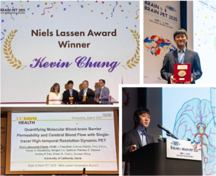
Congratulations to Dr. Kevin Chung for receiving the Niels Lassen Award at Brain & Brain PET 2025! This prestigious award is presented by the International Society for Cerebral Blood Flow and Metabolism to recognize an outstanding scientific contribution made by a young scientist at the brain meeting.
More details of this award-winning work can be found in his recent two publications in the Journal of Nuclear Medicine and Nature Communications. This recognition highlights Kevin’s innovative contributions to brain PET imaging. His work is helping push the frontier of quantitative neuroimaging with total-body PET.
Kevin is jointly mentored by Dr. Guobao Wang, Professor and Associate Vice Chair of Research in the Department of Radiology, and Dr. Simon Cherry, Distinguished Research Professor in the Departments of Biomedical Engineering and Radiology.
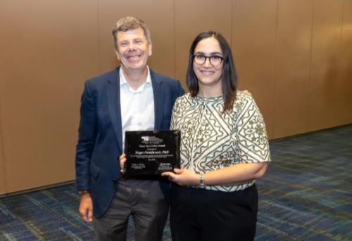
June 2025 – Ramsey Badawi Awarded the 2025 Hibbard-Williams Award
We are proud to share that Professor Ramsey Badawi, Co-Director of the EXPLORER Molecular Imaging Center, has been awarded the 2025 Hibbard Williams Extraordinary Achievement Award by the University of California, Davis - School of Medicine, recognizing his outstanding contributions to research, education, and service.
Dr. Badawi is internationally known for his pioneering work in molecular imaging and PET instrumentation, and is the co-developer of the EXPLORER total-body PET scanner—the world’s first system capable of imaging the entire body simultaneously with unprecedented sensitivity and speed. His visionary leadership has placed University of California, Davis at the forefront of total-body imaging and continues to shape the future of biomedical research.
Please join us in congratulating Dr. Badawi on this well-deserved recognition!
Courtesy of UC Davis Health Radiology.
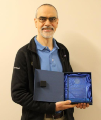
June 2025 – Clemens Mingels awarded the Alavi-Mandell Publication Award for his important work in pediatric PET imaging
Congratulations to Dr. Clemens Mingels, Assistant Attending Physician at Inselspital University Hospital of Bern, for being awarded the Alavi-Mandell Publication Award by the Society of Nuclear Medicine and Molecular Imaging (SNMMI)! This work on “Dose Reduction in Pediatric Oncology Patients with Delayed Total-Body [18F]FDG PET/CT”, published in the Journal of Nuclear Medicine in 2024, was conducted while he was a postdoctoral scholar in Dr. Lorenzo Nardo’s lab at the EXPLORER Molecular Imaging Center.
The Alavi-Mandell Publication Award is given to individuals who were the first author of a paper published in the Journal of Nuclear Medicine who were trainees at the time of publication and made a major contribution to the completion of the work. Congratulations to Dr. Mingels and the co-authors!
May 2025 – EXPLORER investigators touted as SNMMI Ones to Watch
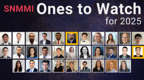
Congratulations to Dr. Brahim Mehadji and Dr. Siqi Li for being recognized as 2025 “Ones to Watch” by the Society of Nuclear Medicine and Molecular Imaging (SNMMI)!
This prestigious honor highlights early-career professionals who are making exceptional contributions to research, education, and clinical practice in nuclear medicine and molecular imaging. Drs. Mehadji and Li are already making waves in the field with their innovative work and dedication to advancing precision diagnostics and therapy.
Thanks to Dr. Emilie Roncali and Dr. Guobao Wang for their nominations. Please join us in congratulating these outstanding junior investigators on this well-deserved recognition!
May 2025 – EXPLORER at the UC Davis Research Expo

The EXPLORER Molecular Imaging Center and the Department of Radiology had another great showing at the UC Davis Research Expo! Thanks to Dr. Benjamin Spencer, Technical Director of the EXPLORER Molecular Imaging Center and Assistant Professor in the Department of Radiology, and Ekatarina Shanina, PhD Candidate in the Department of Biomedical Engineering, for representing our Department and Center!
April 2025 - New Technique Enables Practical Parametric PET Imaging from Clinical Total-Body Scans
Assistant Project Scientist Siqi Li has developed a practical method for generating high-quality parametric images from clinical total-body scans without increasing scan time and costs. The approach, called the relative Patlak (RP) plot, eliminates the need for early-time input data traditionally required by the standard Patlak model. Combined with a self-supervised deep-kernel noise reduction method, the technique allows accurate tracer kinetics to be derived from short clinical scans.
In a study involving 22 human subjects, the RP method produced results closely matching the standard 1-hour Patlak analysis, while improving lesion contrast and even enabling detailed visualization of the heart. Clinical application to 20-minute scans from cancer patients further demonstrated its potential for routine use.
This breakthrough significantly enhances the practicality of total-body PET for dynamic imaging, opening the door to faster, more accessible, and potentially more informative scans in both research and clinical settings.
Related Link:
- Publication - https://jnm.snmjournals.org/content/66/4/654
March 2025 - COBRA 2025 Unites Bay Area Nuclear Imaging Community at UC Davis
March 4, 2025 – Sacramento, CA
This year’s COBRA (Community Of Bay area RAdionuclide imagers) meeting brought together a diverse group of researchers, trainees, and professionals in nuclear imaging from across Northern California for a day of scientific exchange and community-building at UC Davis’ Betty Irene Moore Hall.
The day kicked off with a workshop on fellowship and grant writing led by Katie Hellier (UC Santa Cruz), followed by a networking lunch.
The afternoon program featured two scientific speaker sessions, showcasing cutting-edge research across the participating institutions, including UC Davis, UC Santa Cruz, UC San Francisco, UC Berkeley, Lawrence Berkeley National Lab, and Stanford University. The presentations covered topics like CNS-targeted radi tracers, LGAD detectors for medical imaging, instrumentation development, innovations in imaging for radiation therapy and ultra- thin silicon tracking for autoradiography.
Participants then enjoyed an informal poster session and coffee break, complemented by a research-themed BINGO game to spark interaction and discovery among colleagues. The meeting concluded with a career-oriented panel discussion moderated by Katie Hellier, featuring professionals from academia, national labs, medical physics, and industry.
The organizers also announced a mini-proposal competition with a $500 award aimed at fostering inter-institutional collaboration and supporting new research ideas. The day concluded with a dinner buffet and cold drinks—another opportunity for attendees to connect and unwind. Inspired by the collaborative spirit and rich exchange of ideas throughout the event, the organizing team announced plans to launch a monthly newsletter to keep participants informed about ongoing research efforts across institutions until the next COBRA gathering.
The COBRA organizing committee thanks all who attended and contributed to the event’s success, as well as our generous sponsors – the UC Davis Department of Radiology and Hamamatsu Photonics. Those interested in helping shape future meetings are encouraged to get involved.
Organizing Committee 2025
- Ekaterina Shanina, Reimund Bayerlein, Daehee Lee – UC Davis
- Kimia Gholami, Katie Hellier – UC Santa Cruz
- Ananya Goyal – Stanford University
- Seungeun Lee – Lawrence Berkeley National Lab
March 2025 – New PET Method Maps Blood-Brain Barrier Transport with High-Speed Imaging
A new study led by Kevin Chung, along with Drs. Guobao Wang and Simon Cherry at UC Davis, presents a cutting-edge PET imaging technique to quantify blood-brain barrier (BBB) permeability in humans. Published in Nature Communications, the research shows that dynamic PET scans with high temporal resolution (1-second and 2-second frames) can noninvasively track how different molecules cross the BBB—without the need for blood sampling or multiple tracers.
Using this fast-imaging framework, the team analyzed three tracers: [18F]FDG, [18F]fluciclovine, and [11C]butanol. Each showed distinct patterns of BBB transport consistent with their biological properties. The method enabled simultaneous estimation of blood flow and molecular transfer rates in just two minutes of scan time.
Importantly, the approach also detected age-related changes and reduced transport in individuals with liver inflammation, suggesting broader clinical applications in studying brain-liver axis.
This novel PET framework offers a rapid, noninvasive tool for mapping BBB function, with potential relevance in aging, neuroinflammation, drug development, and metabolic disorders.
Congratulations to Chung et al. and the UC Davis team on this important breakthrough!
Related link:
- In SNMMI newsletter
- Full story in Medical Xpress
January 2025 - Exceptional Quantitative Performance of NeuroEXPLORER Brain PET Scanner
Assistant Project Scientist Negar Omidvari and her colleagues have demonstrated the high quantitative precision and accuracy of the NeuroEXPLORER, a next-generation dedicated brain PET system. The paper, now available in the Journal of Nuclear Medicine, presents extensive evaluations using phantom and human data across a broad range of imaging conditions relevant to dynamic neuroimaging.
The NeuroEXPLORER’s extended axial field of view, combined with its high spatial resolution and sensitivity, was shown to deliver consistent quantitative performance—even with low activity levels and short frame durations. Key findings include less than 5% bias across a wide activity range and sub-1% variations in r covery coefficients, confirming the scanner’s suitability for high-resolution dynamic brain studies.
These results underscore the NeuroEXPLORER’s potential to advance research in neurological disorders by enabling more precise, flexible, and sensitive molecular imaging.
Related Link:
- Publication - https://jnm.snmjournals.org/content/66/1/150.abstract
December 2024 - Affordable PET Imaging Moves Closer with Innovative Plastic-Based Scanner
In an international collaboration with Jagiellonian University in Kraków, Poland, a data correction framework was developed for the modular J-PET scanner—a cost-effective PET imaging system that uses long plastic scintillators instead of traditional, expensive inorganic crystals. Manish Das, a PhD scholar from the J-PET team and UC Davis-based Assistant Project Scientist Reimund Bayerlein successfully demonstrated promising image quality using phantom data for the first time.
The study focused on developing key data correction techniques, including attenuation correction, random coincidence correction using a delayed time window, and scatter correction via Monte Carlo simulations. A test using the standard NEMA image quality phantom showed that these methods significantly improved image uniformity and reduced unwanted residual activity. While the J-PET system still faces challenges in sensitivity and resolution, the results mark a major step toward affordable PET scanning with long axial field of view. The team aims to extend their corrections to human imaging, potentially expanding access to advanced metabolic imaging worldwide.
Related Link:
October 2024 – Development of a Versatile PET Phantom for Realistic Brain Imaging Simulations
PhD candidate Ekaterina Shanina and her colleagues have introduced a groundbreaking phantom technique for positron emission tomography (PET), enabling the simulation of complex static and dynamic activity distributions from a single PET scan.
Using the uEXPLORER total-body PET/CT scanner and a 22Na point source mounted on a robotic arm, the team collected a high-statistics dataset and subsampled it to create arbitrary activity distributions, such as a simple geometric phantom, a high-resolution FDG brain phantom, and even multiple independent frames of florbetaben distribution, a tracer used to image beta-amyloid plaques in the brain. The technique demonstrated strong quantitative accuracy, with errors under 5% across a range of activity concentrations and tissue types.
Unlike conventional phantoms, this universal method offers unmatched flexibility for evaluating image reconstruction, correction techniques, and PET system performance—particularly for the development of next-generation high-resolution brain PET scanners.
Related Link:
August 2024 — Brown Fat Found to Help Clear Harmful Amino Acids Linked to Obesity and Diabetes
A new study led by Yasser Abdelhafez, Maria Chondronikola, and a team at UC Davis reveals that brown adipose tissue (BAT) — also known as brown fat — plays a previously underappreciated role in removing branched-chain amino acids (BCAAs) from the bloodstream. These amino acids, while essential for the body, are often elevated in people with obesity and type 2 diabetes, where they are linked to insulin resistance and poor metabolic health.
Published in iScience, this research utilized advanced 18F-fluciclovine total-body PET/CT imaging to track how the body processes BCAAs. The team found that the supraclavicular region (where brown fat is most active in humans) had a higher uptake of fluciclovine — a BCAA analog — compared to other fat depots like abdominal or chest fat. Interestingly, individuals with obesity or diabetes showed lower BCAA clearance in brown fat, suggesting reduced metabolic activity. The study also analyzed fat biopsies and found that people with high BAT volume expressed more genes related to BCAA breakdown, further confirming brown fat’s role in amino acid metabolism.
These findings suggest that BAT could serve as a natural “sink” for clearing excess BCAAs, making it a potential therapeutic target for improving metabolic health.
Related Link:
March 2025 – Total-Body PET Improves Nodal Staging Accuracy in Lung Cancer Patients
A new study led by Clemens Mingels at UC Davis reveals that Total-Body (TB) PET/CT imaging offers higher diagnostic accuracy than conventional PET/CT in staging non-small cell lung cancer (NSCLC). Published in the European Journal of Nuclear Medicine and Molecular Imaging, the prospective head-to-head comparison assessed 48 patients, imaged with TB PET/CT and short-axial field-of-view (SAFOV) PET/CT, both performed on the same day, using the same radiotracer, [18F]FDG.
The findings showed that TB PET/CT provided slightly higher sensitivity (86.0% vs. 77.2%) and positive predictive value, with fewer staging errors compared to SAFOV PET/CT. Importantly, TB PET/CT detected mediastinal lymph node metastases at lower tumor-to-background ratios and smaller volumes, suggesting improved ability to identify small, low-uptake lesions.
Congratulations, Clemens et al.!
Related Link:
December 2024 – A new study led by Drs. Yasser Abdelhafez, Siba Raychaudhuri and Abhijit Chaudhari at UC Davis Health has demonstrated the value of Total-Body PET imaging in identifying systemic inflammation in patients with psoriatic and other autoimmune arthritis.
The research, published in Rheumatology included 71 adults with psoriatic arthritis, rheumatoid arthritis, or osteoarthritis, and revealed that a significant proportion of inflamed joints (15%), entheses (20%), and nails (13%) were undetected by routine assessments but captured by a single, low-dose Total-Body PET scan. This work could pave the way for earlier diagnosis and better treatment of autoimmune arthritic conditions.
Congratulations to the UC Davis team!
Related Link:
- Publication - https://academic.oup.com/rheumatology/advance-article/doi/10.1093/rheumatology/keae702/7931525
March 2025 - Negar Omidvari Honored with Excellence in Research Award
Dr. Negar Omidvari, Assistant Project Scientist in the Department of Biomedical Engineering, has been recognized with the UC Davis Academic Federation Excellence in Research Award for her groundbreaking contributions to total-body PET imaging. Negar’s research focuses on using dynamic PET imaging to study the immune system, including mapping T-cell distribution in the human body. Her work has provided insights into immune response and memory, particularly in COVID-19 patients. She was also the first to apply a CD8-targeted radiotracer in this context, contributing to advancements in infectious disease imaging. Her findings were published in Science Advances.
Congratulations, Negar, on this achievement!
Related Link:
February 2025 - Siqi Li Awarded NIH R03 Grant for Advancing Total-Body PET Parametric Imaging
Dr. Siqi Li, Assistant Project Scientist, Department of Radiology, has been awarded a two-year NIH/NIBIB R03 research grant as a Principal Investigator to explore the feasibility of Total-body PET Parametric Imaging using the Relative Patlak Plot. His work aims to refine quantitative imaging techniques that enhance the efficiency and accuracy of parametric PET imaging, paving the way for improved disease detection and monitoring.
Siqi’s method builds on his recent JNM paper, introducing a streamlined approach to total-body dynamic PET imaging that eliminates the need for early-time input functions, making it more practical for clinical applications. The project will be conducted at the UC Davis Department of Radiology, with funding totaling $161,000 for two years from the National Institute of Biomedical Imaging and Bioengineering (NIBIB). This grant marks an exciting milestone in Siqi’s research career and represents a step forward in developing innovative PET imaging methods.
Congratulations, Siqi, on your first grant!
Related Link:
- Publication - https://jnm.snmjournals.org/content/jnumed/early/2025/02/27/jnumed.124.268496.full.pdf
December 2024 - Kevin Chung Awarded Prestigious AHA Postdoctoral Fellowship

The American Heart Association (AHA) has awarded Dr. Kevin Chung a highly competitive two-year Postdoctoral Fellowship to support his research on multiparametric PET imaging of the heart-brain axis. His project aims to develop a methodological framework using total-body dynamic 18F-FDG PET to assess vascular and metabolic function in both the heart and brain, a crucial step in understanding multisystemic effects of myocardial ischemia.
Kevin, who is jointly supervised by Dr. Guobao Wang and Dr. Simon Cherry, will collaborate with a mentoring team that includes cardiologist Dr. Javier Lopez and neuroimaging scientist Dr. Audrey Fan. His fellowship, totaling $161,248, will span from January 2025 to December 2026. Through his research, Kevin hopes to bridge heart and brain imaging, equipping himself with technical expertise in molecular imaging and laying the groundwork for future innovations in inter-organ imaging and pathophysiology research.
Congratulations, Kevin!
Related Link:
- Wang Lab - https://wanglab.faculty.ucdavis.edu/archives/2886
- Award Detail - https://proposalcentral.com/Insights/yK3zgRfRQcI=/Public/AwardDetails/1360296
October 2024 - Reflections from the 2024 IEEE MIC Conference in Tampa, FL
The 2024 IEEE Nuclear Science Symposium (NSS) and Medical Imaging Conference (MIC) held from October 26 to November 2 in sunny Tampa, FL, brought together some of the brightest minds in medical imaging science to explore the latest developments and to foster collaboration. With plenary presentations by Richard E Carson spotlighting “biomathematics in PET science” and by Sana Karam highlighting the “role of imaging in streamlining treatment regimens”, the event set an inspiring tone for a week of discovery and connection.
Exploring Hot Topics in Medical Imaging
Deep learning continued to dominate the conversation, with exciting advancements showcased in image denoising, data corrections, PET image reconstruction or multi-modal nuclear medicine imaging. Workshops and short courses provided valuable opportunities for hands-on learning in areas such as kinetic modeling, AI-powered image processing, and rapid PET image reconstruction. Also of interest were the GATE and CASToR user meetings, where attendees exchanged insights on simulation and image analysis platforms. And anybody working on detector hardware and signal read-out was given the opportunity to have a fruitful exchange with representatives of manufacturing companies at the industry exhibition.
UC Davis Shines on the Global Stage
UC Davis researchers from the Departments of Radiology and Biomedical Engineering made a strong showing at MIC 2024, presenting an impressive collection of posters and oral presentations. Our contributions spanned diverse topics, including novel image reconstruction methods, quantitative image quality assessment, kinetic modeling, deep learning-based reconstructions, innovative phantom designs, and advancements in detector development and characterization. These efforts highlighted the depth and breadth of expertise within our team and sparked engaging discussions with peers from around the globe.
Building International Connections
The conference also provided a fantastic opportunity to strengthen international collaborations. Our research group, with established partnerships with institutions all over the world, was able to meet with collaborators face-to-face, reinforcing these vital relationships. And who knows who many novel and exciting ideas have emerged during countless in-depth discussions over lunch or dinner?
Memorable Moments and Celebrations
Networking and relaxation were equally well-supported by MIC’s vibrant social events. The Welcome Reception delighted attendees with an array of delicious food, beverages, and conversation, setting the stage for reconnecting with old friends and meeting new ones. The MIC Dinner, a picturesque three-hour boat cruise through Tampa Bay, was another standout experience, offering live music, dancing, and a perfect close to the week.
Recognizing Excellence
We are thrilled to celebrate two exceptional UC Davis researchers who earned top honors at MIC 2024.

Xiaoyu Duan secured second place for presentation of her innovative work on “Quantification of 18F-FDG Delivery Rate for Liver Inflammation using Shortened Dynamic PET Imaging Protocols.” Her work was recognized as a valuable contribution to the field of kinetic modeling and parametric imaging showcasing potential clinical applications.
Ekaterina Shanina took home the first-place prize for her presentation “A Universal Activity Painting Phantom for Positron Emission Tomography.” Her cleverly named PICASSO phantom, inspired by the painter’s light-drawing techniques, captivated the audience with its creativity and countless applications.
Looking Ahead
MIC 2024 proved to be an inspiring blend of cutting-edge science, meaningful collaboration, and community celebration. We leave Tampa motivated and with new ideas and stronger connections moving forward. Many attendees are already looking forward to next year’s conference taking place in Yokohama, Japan.
---
Authored by Reimund Bayerlein
11/15/2024
November 2024 - EMIC Diwali Celebration
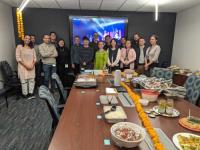
Explorer Molecular Imaging Center (EMIC) Celebrated Diwali (also know as Deepawali/Tihar in Southeast Asia) festival on November 8, 2024, by means of a team potluck. Shivani and Atul led the organizational effort and we enjoyed dishes such as pulao rice, jalabi, gulab jamon, onion bhajis, home-baked naan and spiced chickpea salad, amongst others.
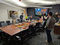
Simon’s chicken and paneer curries (made in the best English style) were a triumph, but Ramsey’s attempt at burfi was definitely a work-in-progress – perhaps next time he will dare to actually bring them in to share! Music and a video from Southeast Asia were showcased during the celebration.
A great time was had by all and we look forward to doing it again next year!
September 2024 - UC Davis scientists present at Total Body PET 2024
September 19, 2024, Simon Cherry, Lorenzo Nardo, Kevin Chung and Quyen Tran will also be presenting at this year’s Total Body PET meeting in Groningen, Netherlands. Read More.
May 2024 - Total body nuclear medicine PET scan continues to evolve
May is National Cancer Research Month, and nuclear scientists at UC Davis are giving doctors a tool to see patients as a whole and not just the part of their bodies making them sick. [ Watch Video ]

November 2018 - Human Images From World's 1st Total-Body Scanner Unveiled
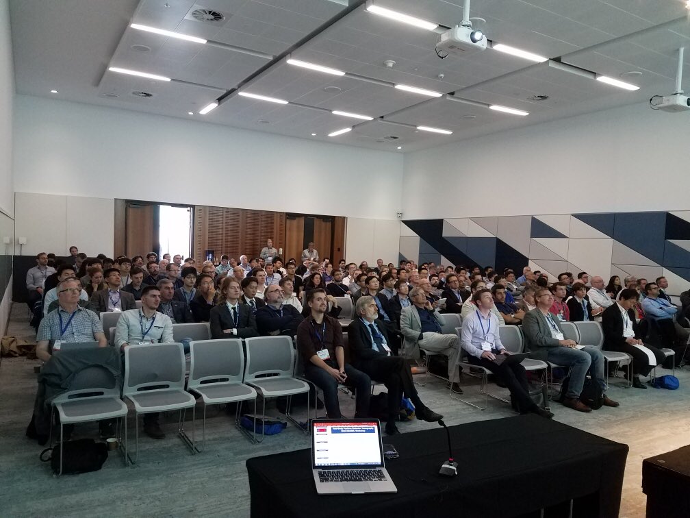
The first human images from the fully completed EXPLORER scanner have been unveiled at the IEEE MIC 2018 Total-Body PET workshop. The work was presented by Dr. Eric Berg in Sydney, Australia. View presentation PDF
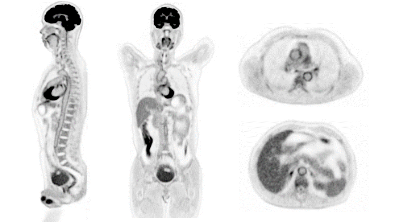
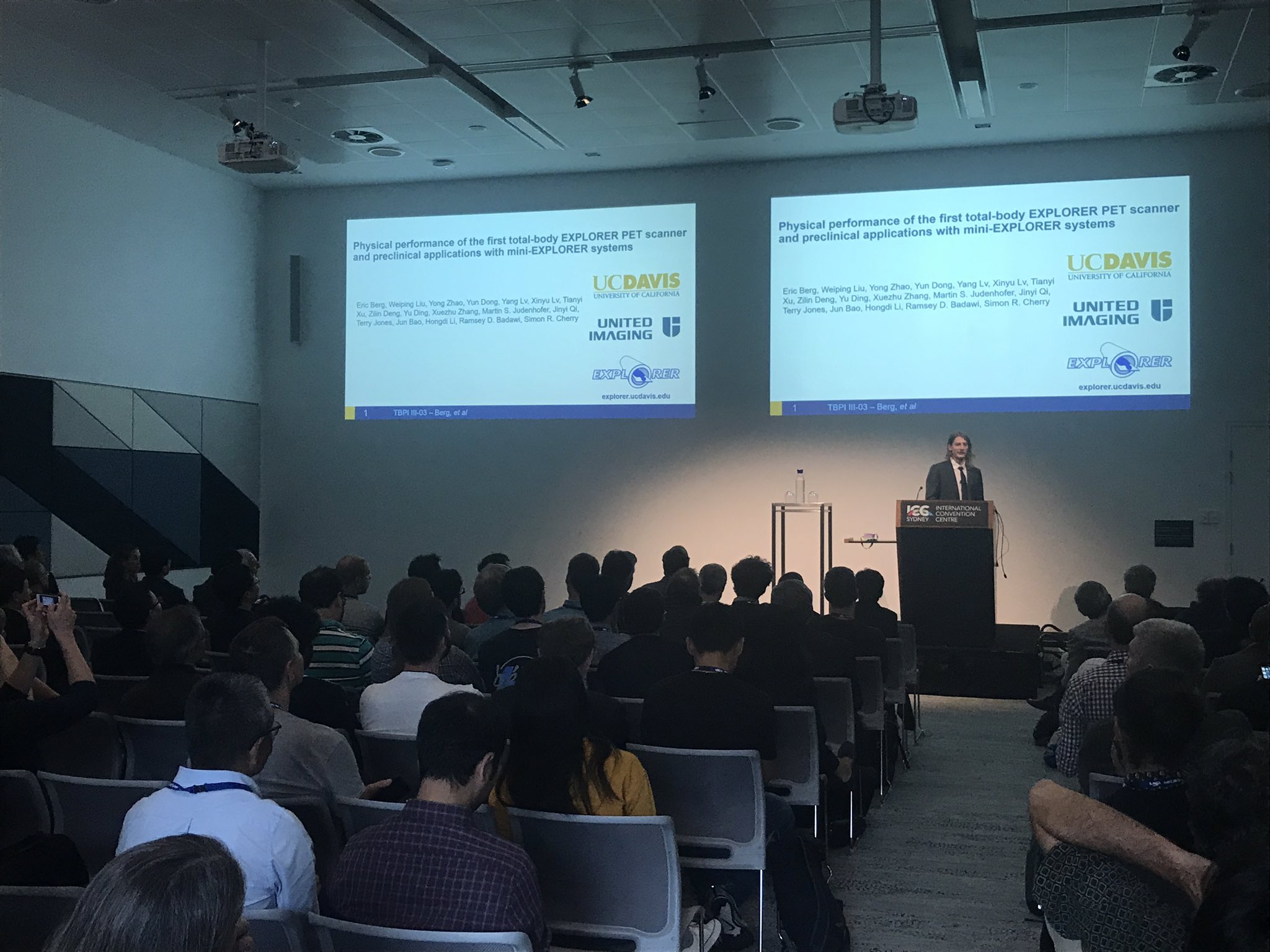
November 2018 - Total-Body PET Workshop (November 17th, 2018, IEEE NSS/MIC, Sydney, Australia)
More than 150 researchers from around the world attended a workshop on total-body PET imaging held in conjunction with the IEEE NSS/MIC Conference in Sydney, Australia. Presentations covered strategies for electronics and detectors, system simulations, and phantom and first human images from the two recently completed scanners, the 194 cm long uEXPLORER scanner by the UC Davis/United Imaging team, and the 70 cm long PennPET EXPLORER scanner from the Penn/KAGE Medical team. Dr. John Sunderland from the University of Iowa also gave an invited talk discussing possible approaches to measuring the performance of these new imaging systems.
November 2018 - Postdoctoral positions available
The EXPLORER program at UC Davis has vacancies for several postdoctoral fellows in topics such as imaging physics, kinetic modeling, image reconstruction and translational imaging. Interested candidates should apply to Drs. Simon Cherry and Ramsey Badawi by December 15th 2018.
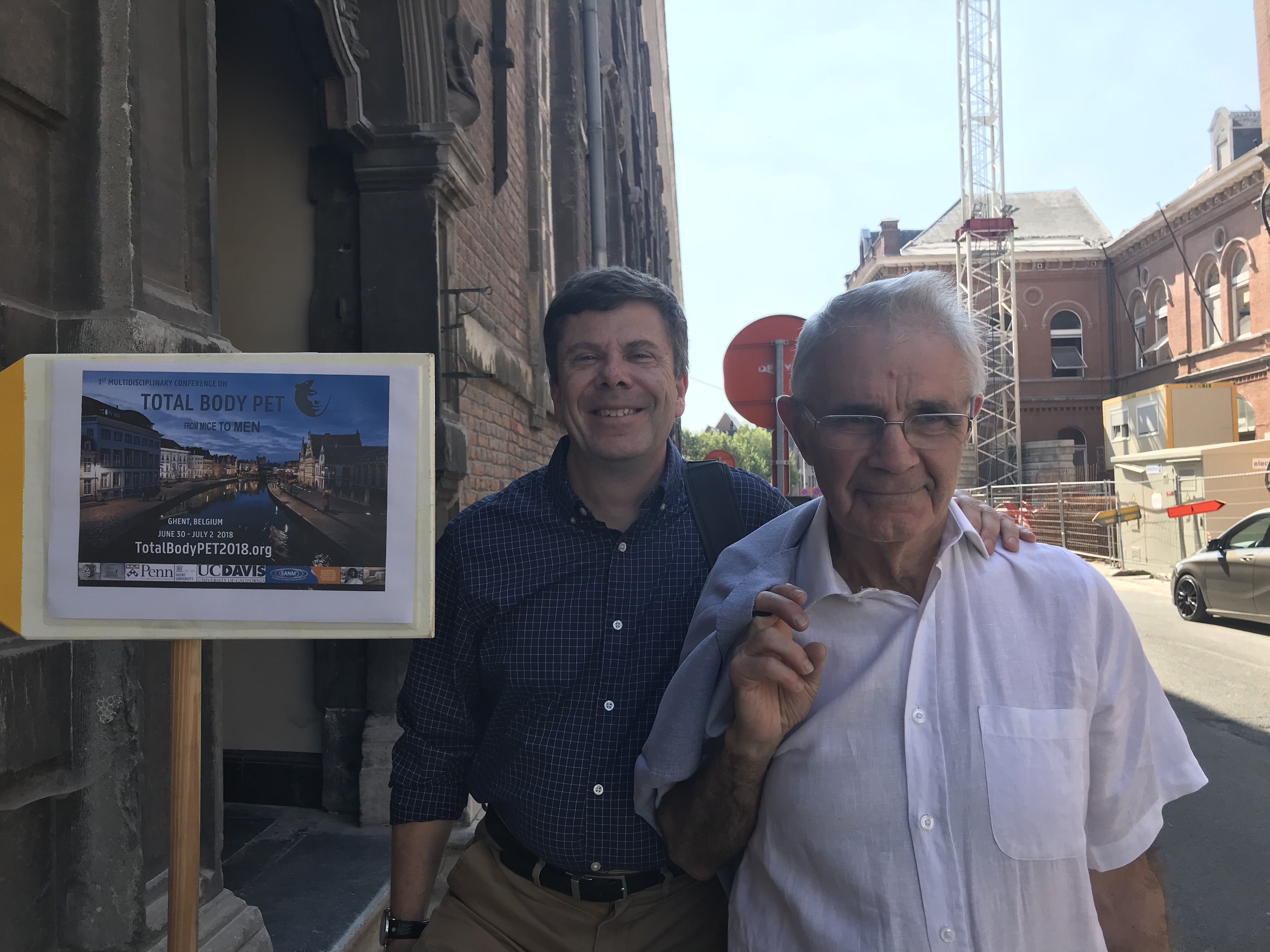
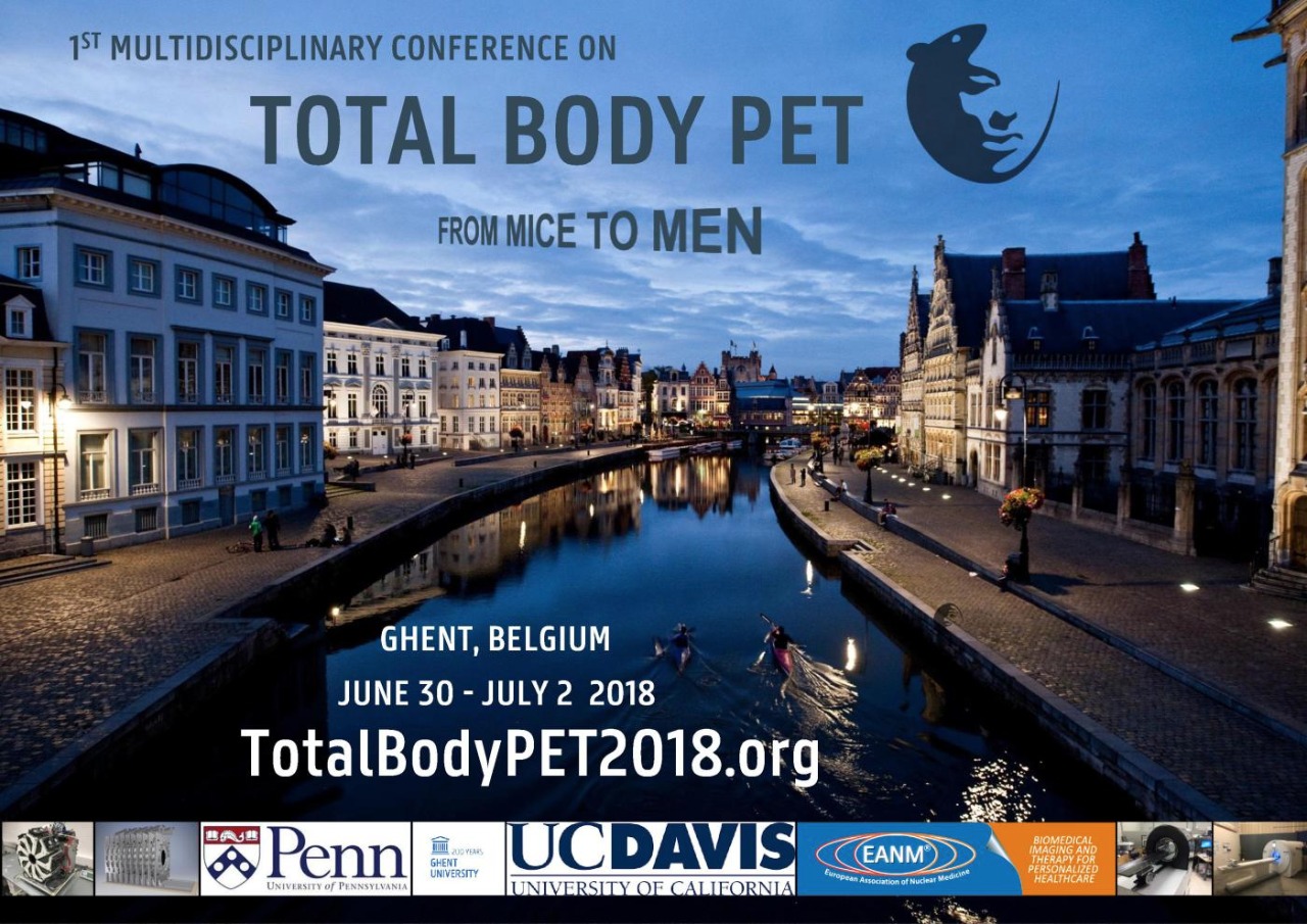
July 2018 - First Conference on Total-Body PET held in Ghent
The first conference on Total-Body PET attracted over 120 participants from across the world (Ghent, Belgium, June 30-July 2, 2018). The conference featured multiple presentations from both the UC Davis and U Penn components of the EXPLORER project. Professor Terry Jones (Radiology, UC Davis) gave a delightful talk on the potential applications of the EXPLORER scanner.
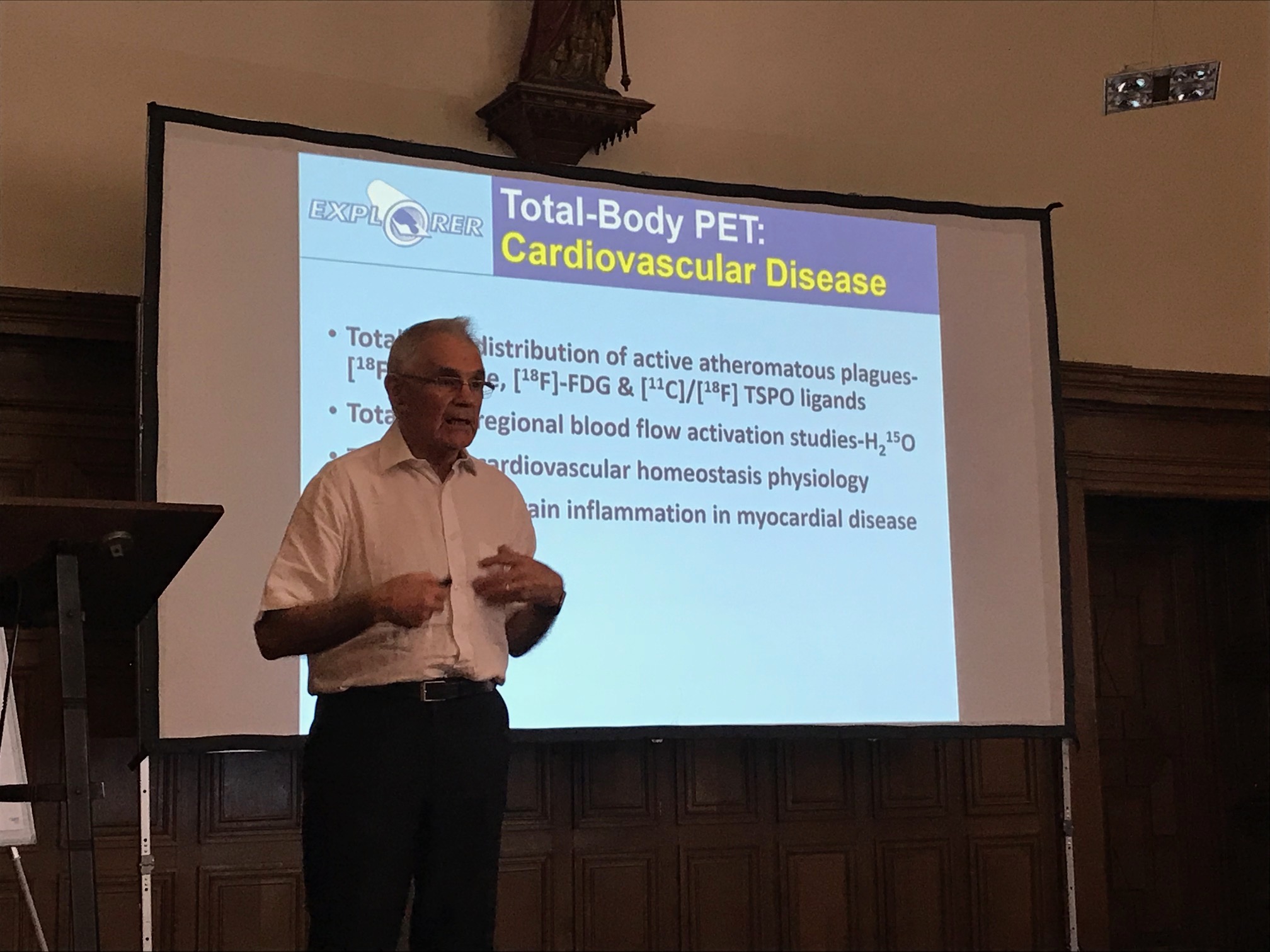
Professors Jones presenting at the Total-Body PET conference
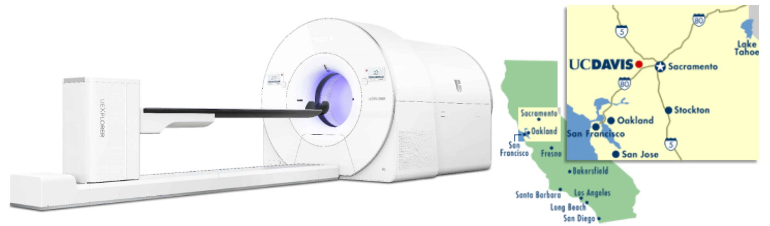
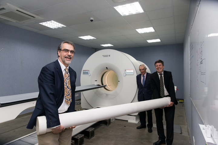
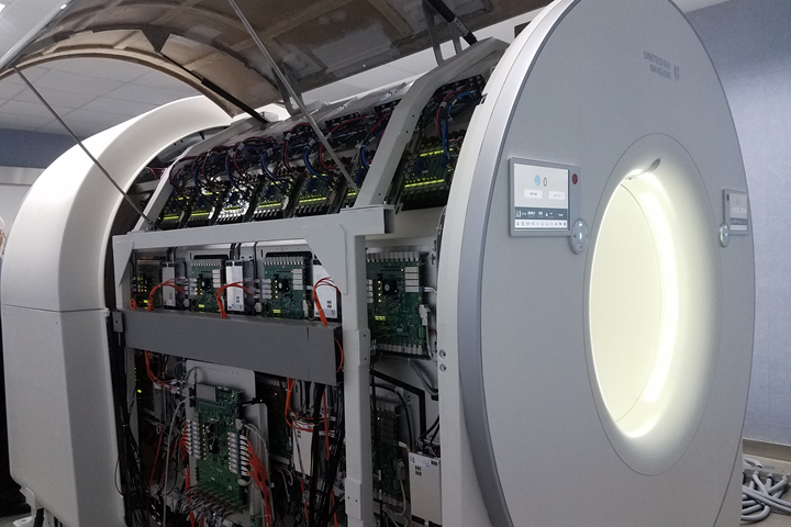
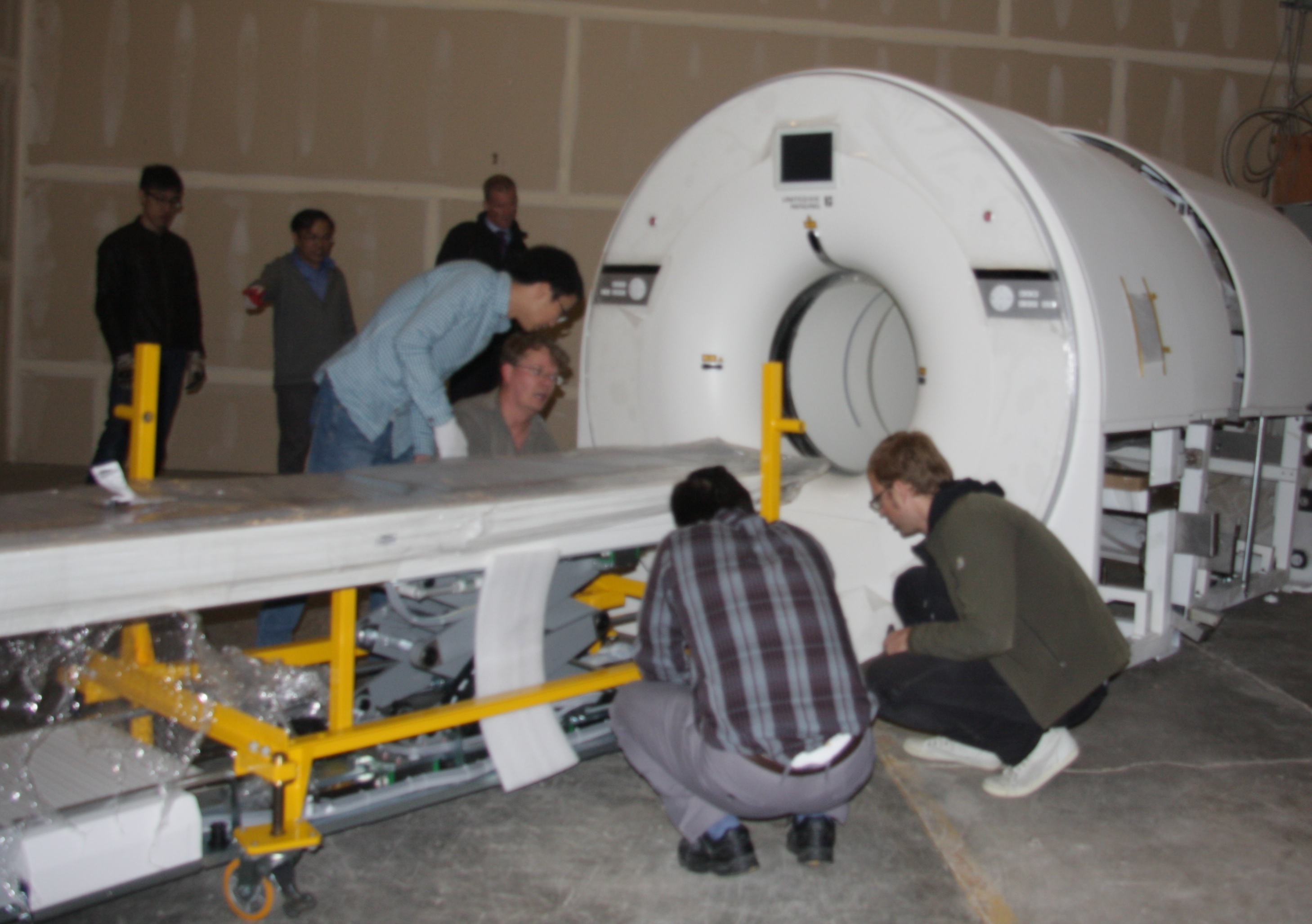
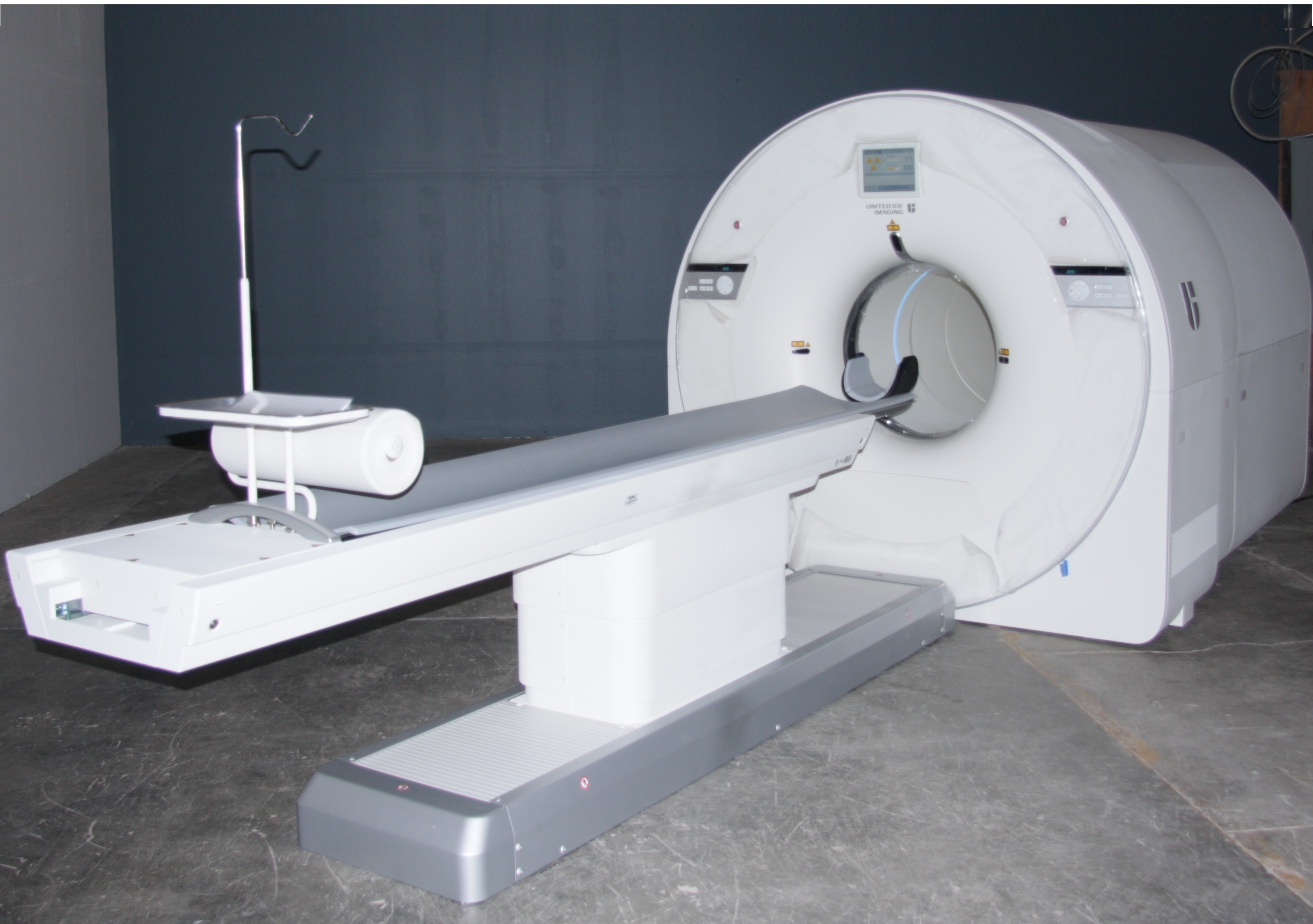
May 2018 - EXPLORER Total-Body PET Scanner Completed
Fabrication of the first ever total-body PET scanner has been completed in collaboration with our industry partners United Imaging Healthcare and SensL Technologies. The scanner has a 194 cm axial field of view for PET imaging provided by over 500 thousand detector elements. It also contains a low dose CT scanner for anatomical imaging and PET attenuation correction. Characterization of the scanner is currently underway with the first ever human imaging set to take place in the near future.
February 2018 - Mini-EXPLORER II
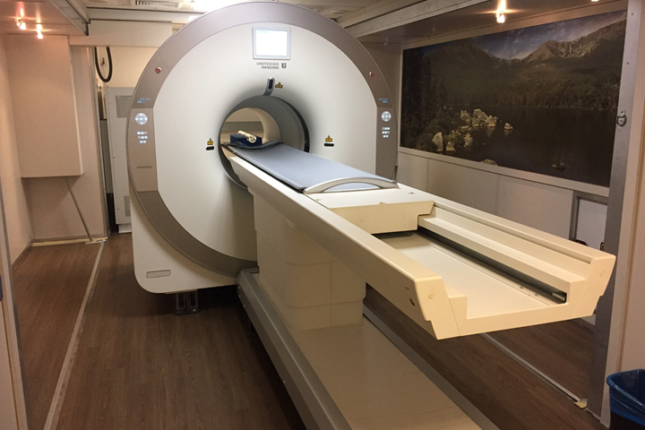
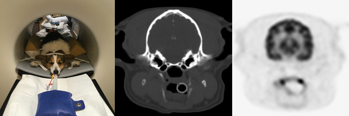
In collaboration with United Imaging Healthcare and the UC Davis department of Veterinary Medicine, the Mini-EXPLORER II small animal PET/CT scanner has been successfully installed and is currently in use at the veterinary hospital. This scanner uses the same detectors and electronics as the full human EXPLORER but has been scaled down for small animal imaging with an axial field of view of 48.4 cm.
November 2017 - Save the date: Total-Body PET - From Mice To Men
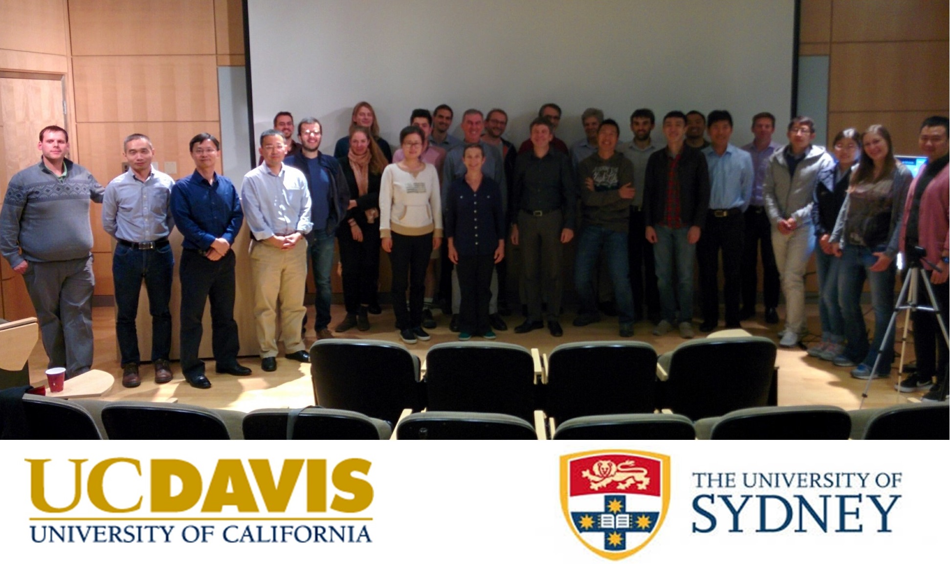
October 2017 - Joint UC Davis - University of Sydney Workshop on EXPLORER Methodology
UC Davis and the University of Sydney held a joint workshop on October 18th, 2017 to discuss methodology and computational imaging in support of the EXPLORER total-body PET scanner project. Presentations covered latest technology developments, the challenges associated with tracking and correcting for subject motion during imaging, methods for reconstructing the massive EXPLORER datasets and ensuring the resulting images are quantitatively correct, and methodology to model time-series of imaging data to extract specific parameters related to radiotracer transport (blood flow), metabolism and binding. More than thirty participants joined the workshop held at UC Davis, including seven visitors from the University of Sydney. The workshop also was webcast to a further 20 participants located in three different countries. This workshop was funded by a joint UC Davis - University of Sydney Priority Partnership Collaboration Award administered by the Office of Research. The next workshop is planned for April 2018 in Sydney.
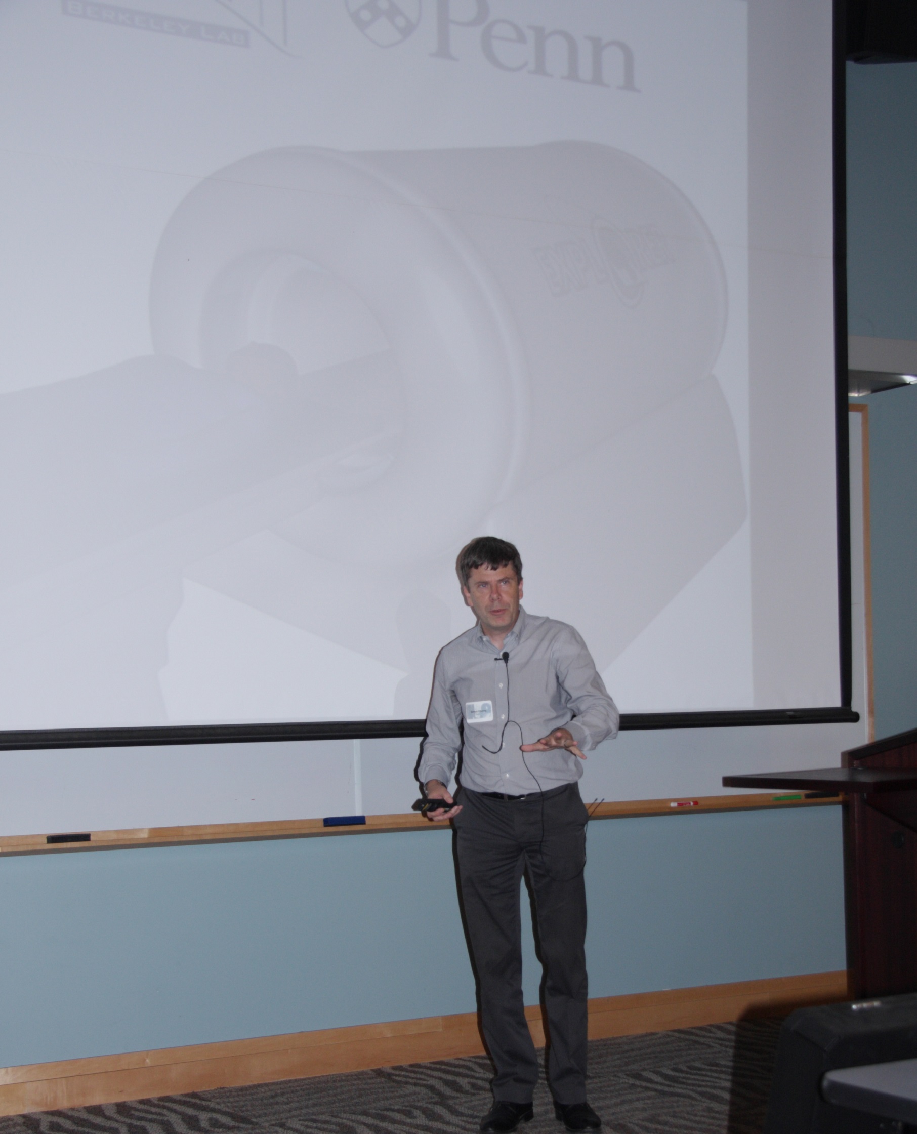
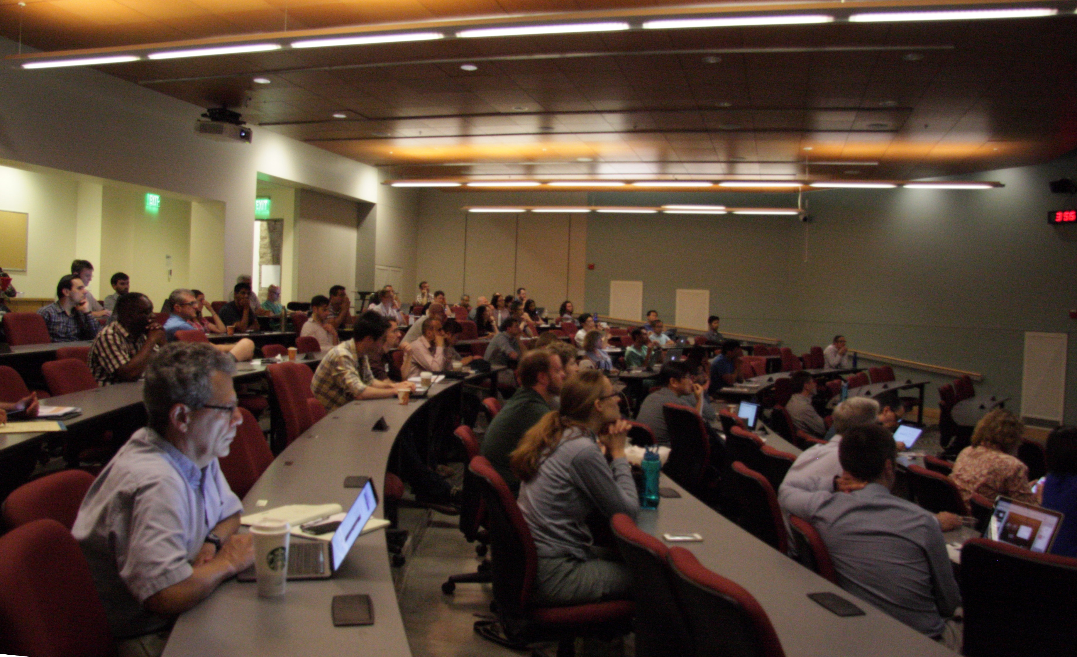
September 2017 - UPenn EXPLORER Workshop 2017
June 2017 - UC Davis miniEXPLORER Symposium
On June 19, UC Davis held the miniEXPLORER symposium at the School of Veterinary Medicine, with an attendance of ~75 people. With the number One Veterinary School in the country, the School of Medicine, the Comprehensive Cancer Center, and the Department of Biomedical Engineering, UC Davis provides a unique environment for translational research. The vision of this symposium was the demonstration of the capabilities of PET for companion and research animals, as well as the power of PET for translational studies. Eight oral presentations showcased studies that have been done with the first miniEXPLORER prototype or will be done with the miniEXPLORER II that we anticipate to be functional in Fall 2017. The event was followed by a reception to promote networking and discussions of potential collaborations between investigators. Incentive for research will be given in the form of four pilot grants to be awarded in September and presented at the second part of the symposium planned in the Fall.
January 2017 - EXPLORER Consortium Selects Industry Partners
The EXPLORER consortium has selected two industry partners to help build the first prototype scanner. They are United Imaging Healthcare (UIH) America, a North American subsidiary of Shanghai United Imaging Healthcare, and SensL Technologies of Cork, Ireland. UIH America was selected to build the first total-body PET scanner after an extensive review of potential commercial partners. "We are very proud to be selected to be part of the EXPLORER consortium and provide the system design support required to deliver a system capable of outstanding performance,"said Hongdi Li, CEO of UIH America. The detector technology in the scanner will incorporate the latest generation solid-state silicon photomultiplier light sensors instead of the photomultiplier tubes used in conventional PET scanners. "The use of silicon photomultiplier technology is rapidly replacing older photomultiplier tube technologies and provides improved resolution for a system of this scale," said Bryan Campbell, CEO of SensL, which will supply the detectors. "We believe we have gathered leaders in the medical imaging field to quickly and cost effectively bring this technology to reality in an exciting and innovative way," said Ramsey Badawi, co-leader of the project.
December 2016 - UC Davis Built mini-EXPLORER System
The team at UC Davis in collaboration with Siemens Medical Solutions have recently built and evaluated a prototype PET scanner for high sensitivity and total-body imaging of non-human primates, called the mini-EXPLORER. The mini-EXPLORER was built from a decommissioned clinical PET scanner, and rearranging the components into a scanner suitable for total-body monkey imaging. The mini-EXPLORER scanner will support research studies at the California National Primate Research Center (CNPRC) and allow the EXPLORER team and collaborators to explore new imaging applications enabled by Total-Body PET. The scanner is expected to be deployed by early 2017. First images of a rat imaged dynamically using F-18[FDG] showed excellent image quality and sensitivity of the PET system.
![Dynamic PET image series of a rat imaged with F-18[FDG]](/sites/g/files/dgvnsk6796/files/inline-images/Mini-EXPLORER-ratimage.png)
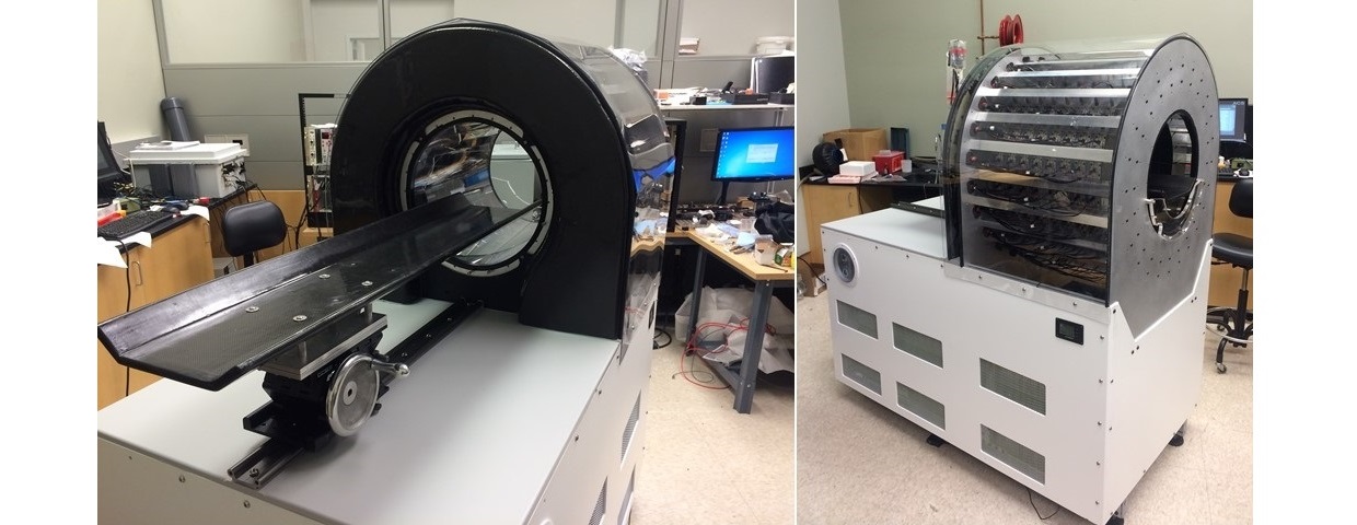
December 2016 - UC Davis Received EXPLORER Mock-up System from UIH America
UC Davis installed a mock-up system of the EXPLORER total-body scanner. The system has the exact same geometry and dimensions as the final system. While the system does not have a functional PET or CT component it provides a fully functional patient bed. This mock up system will be used to investigate patient comfort and develop EXPLORER specific imaging workflows.
November 2016 - IEEE MIC Strasbourg
Experiments on the Feasibility Evaluation of a long axial field-of-view PET scanner for non-human primates have been presented at the IEEE Medical Imaging Conference 2016 in Strasbourg, France.
September 2015 - NIH Transformative Research Award - $15.5 million
Simon Cherry and Ramsey Badawi have received a $15.5 million, 5-year NIH Transformative Research Award to lead the consortium that is tasked with the development of EXPLORER, the world's first Total-Body PET scanner (R01 CA206187).