Positron Emission Tomography – Imaging on Molecular Level
Positron Emission Tomography (PET) is a powerful molecular imaging technique that allows for the 3-dimensional visualization of physiologic, metabolic and molecular processes in the body, making it invaluable in both clinical and research settings. A radio-chemical substance is administered (typically intravenously) into the patient, the distribution of which can be measured with the PET scanner and turned into medical images using mathematical algorithms and data processing. There are thousands of PET scanners across the world performing millions of scans in patients each year.
EXPLORER: Revolutionizing PET Imaging

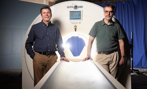
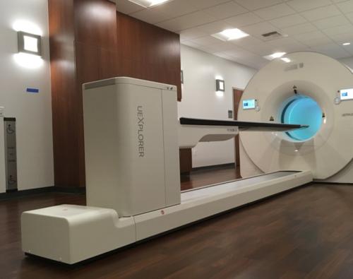
The uEXPLORER scanner, the first clinically approved total-body PET system in the world, was installed in 2019 and represents a significant advancement in this field. In contrast to conventional PET scanners which operate in a step-and-shoot manner imaging 12-30 cm of the human body at a time, the total-body PET scanner is the first medical imaging device capable of total-body simultaneous imaging. This total-body scanner provides drastically higher sensitivity compared to conventional PET scanners. This enhanced sensitivity opens new possibilities for PET applications in biomedical research and clinical practice. The uEXPLORER can achieve improved image quality while scanning faster, using lower radioactive doses, or scanning later after injection.

Before the advent of total-body PET, imaging required the patient to be scanned sequentially in multiple bed positions, which significantly limited the efficiency of detecting emitted signals and reduced the ability to accurately track the dynamics of radiotracer distribution across the entire body. With uEXPLORER, total-body imaging is now feasible without moving the patient, thereby enhancing clinical and research capabilities.
Ongoing and Possible Applications of EXPLORER:
- Improved cancer detection through enhanced contrast and resolution.
- Accelerated imaging for children at low doses, reducing the need for anesthesia.
- Studies of metabolic disorders, autoimmune diseases (such as rheumatoid arthritis), neurodegenerative diseases (such as Alzheimer’s disease), infectious diseases (such as HIV or COVID 19), cardiac diseases, peripheral vascular diseases, and other chronic conditions.
- Conducting low-dose evaluations of new drug pharmacokinetics across all organs.
The uEXPLORER scanner constitutes a paradigm shift in PET imaging, offering numerous advantages and benefits that can enhance standard-of-care and advance nuclear medicine research. Currently, the uEXPLORER at UC Davis performs an average of about 40 high-quality PET scans per week. Our goal is to make total-body PET scanning available to the broader imaging and clinical research communities and for a wide range of imaging applications.
